You are here:
Normal heart case 2
1234567
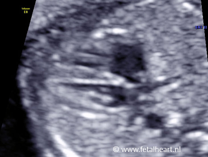
Still of the 4 chamber view.
Size of the heart is normal.
Cardiax axis approximately 45°.
Atria and ventricles are symmetrical.
Pulmonary venous return to left atrium visible.
In this image the crux is clearly visible.
Size of the heart is normal.
Cardiax axis approximately 45°.
Atria and ventricles are symmetrical.
Pulmonary venous return to left atrium visible.
In this image the crux is clearly visible.
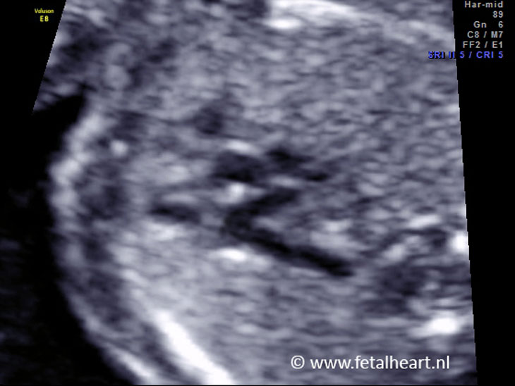
Color Doppler recording of the 4 chamber view.
Proper filling of the ventricles.
No flow across the ventricular septum.
Proper filling of the ventricles.
No flow across the ventricular septum.
Left ventricular outflow tract.
Aortic valve is clearly visible.
The valve is thin and opens smoothly.
Normal sized annulus.
Note how far the aorta the runs rightwards (please compare with the transposition cases).
Aortic valve is clearly visible.
The valve is thin and opens smoothly.
Normal sized annulus.
Note how far the aorta the runs rightwards (please compare with the transposition cases).
Still of the 3 vessel view. P
ulmonary trunk a bit blurred in this picture, though the lateral branching is clearly visible.
ulmonary trunk a bit blurred in this picture, though the lateral branching is clearly visible.
Aortic arch in longitudinal plane.
The aorta arises centrally from the heart and is circular shaped, also called umbrella sign.
The neck vessels arise from the aortic arch.
The aorta arises centrally from the heart and is circular shaped, also called umbrella sign.
The neck vessels arise from the aortic arch.
Inferior and superior caval vein entering the right atrium, this is called bull’s horn sign.
De umbilical vein and ductus venosus is clearly visible in this clip.
De umbilical vein and ductus venosus is clearly visible in this clip.
The ductal arch arises anteriorly from the heart, directly behind the sternum.
The shape resembles an ice hockey stick (less circular than the aorta).
The last part of the aortic arch (with a visible neck vessels) merges with the duct.
The shape resembles an ice hockey stick (less circular than the aorta).
The last part of the aortic arch (with a visible neck vessels) merges with the duct.