You are here:
Hypoplastic aortic arch case 1
1234567891011
Images made at 18 and 28 weeks.
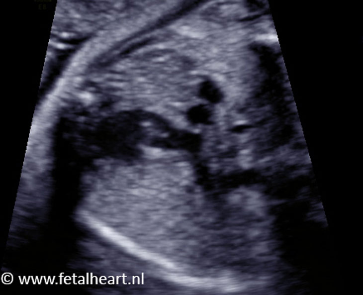
4 chamber view at 18 weeks’gestational age.
Right ventricle is larger than the left ventricle, visible directly beneath the AV-valves.
The left ventricle is however long enough, reaching the apex.
Color Doppler is switched on, no signs of a ventricular septum defect.
The flow across the foramen ovale is abnormal, like a whirlpool with predominantly a direction towards the right atrium (red).
B mode clip of 4chamber view and the annulus of the aortic valve.
Most prominent in this clip is the asymmetry of the ventricles, with a wide right, and small – but long enough – left ventricle.
Atrial septum appears thickened in this projection.
Most prominent in this clip is the asymmetry of the ventricles, with a wide right, and small – but long enough – left ventricle.
Atrial septum appears thickened in this projection.
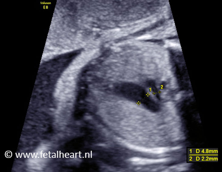
Aorta outflow tract.
Aorta in alignment with the ventricle septum, without signs of a subaortic ventricle septum defect.
The aortic annulus is slightly smaller than normal, but the valve opens normally.
The leaflets are thin and hardly visible, which is normal.
Aorta in alignment with the ventricle septum, without signs of a subaortic ventricle septum defect.
The aortic annulus is slightly smaller than normal, but the valve opens normally.
The leaflets are thin and hardly visible, which is normal.
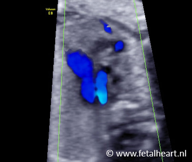
However, with color doppler a small jet is visible just beneath the valve.
Blue and red flow is visible across the small VSD, thus blood is traveling from the right to the left ventricle and vice versa.
This is a normal phenomenon in fetal VSDs.
Blue and red flow is visible across the small VSD, thus blood is traveling from the right to the left ventricle and vice versa.
This is a normal phenomenon in fetal VSDs.
A second clip of the VSD. In slow motion the blood moving back and forth is clearly visible.
Three vessel view. The aorta is too small and therewith positioned too dorsally.
Three vessel trachea view (also known as 3VTV).
Evident difference in size between the ductal and aortic arch.
Aortic arch is too small for a considerable segment.
Evident difference in size between the ductal and aortic arch.
Aortic arch is too small for a considerable segment.
Three vessel trachea view with color Doppler.
Equal direction of flow.
Acceleration si visible in the aortic arch.
Equal direction of flow.
Acceleration si visible in the aortic arch.
Aortic arch with color Doppler.
The arch is tiny and becomes even smaller after the neck vessels.
The arch is tiny and becomes even smaller after the neck vessels.
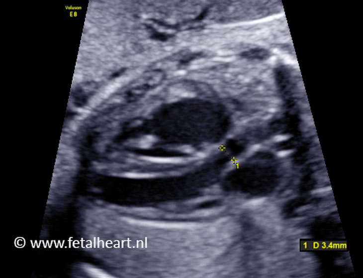
Image of the measurement of the aortic valve.
3.4 mm is too small for 28 weeks’ gestational age.
3.4 mm is too small for 28 weeks’ gestational age.
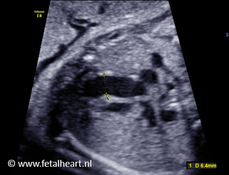
Image of the measurement of the pulmonary valve.
6.4 mm is a normal size for 28 weeks’ gestational age.
6.4 mm is a normal size for 28 weeks’ gestational age.