You are here:
Heterotaxia case 1
1234567891011
Sweep from head to abdomen.
Fetus in cephalic position.
Apex of the heart points to the left side of the fetus.
Stomach at the right side.
The descending aorta is positioned in mid-front of the spine instead of left of the spine.
Fetus in cephalic position.
Apex of the heart points to the left side of the fetus.
Stomach at the right side.
The descending aorta is positioned in mid-front of the spine instead of left of the spine.
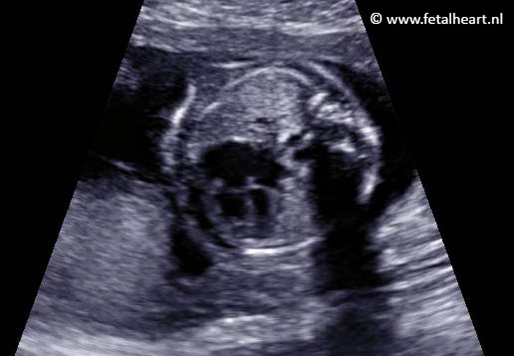
4 chamber view appears quite normal.
The ventricles are a bit asymmetrical: right is larger than left.
The right ventricle is the morphological right ventricle, because the moderatorband is visible in this ventricle.
The left ventricle had features of a morphological left ventricle: the septum is smooth and it forms the apex in a sharp triangle.
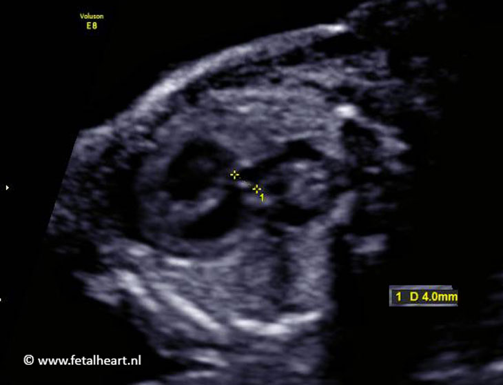
At least 2 VSDs: one directly beneath the AV-valves.
One a bit lower, muscular VSD.
One a bit lower, muscular VSD.
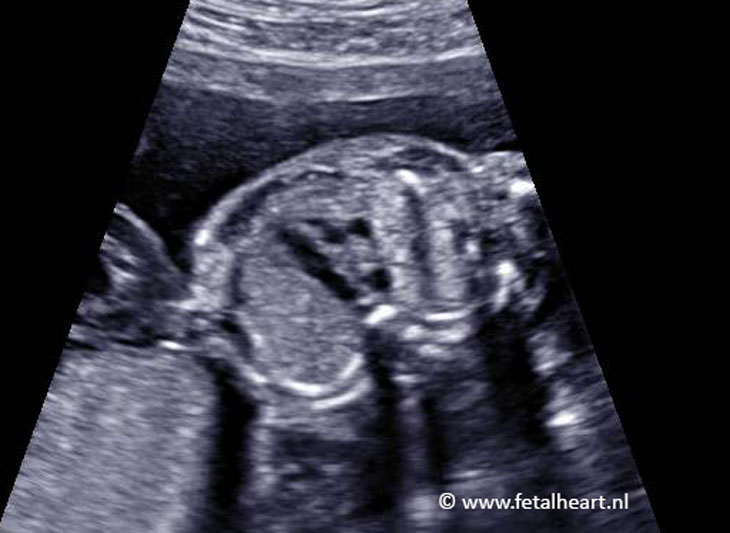
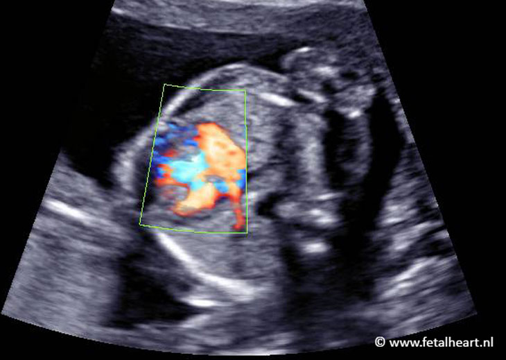
Normal right pulmonary vein entering the left atrium (blue signal).
Long venen van linker en rechter long monden normaal in, in linker atrium.
Bij heterotaxie kunnen er afwijkingen zijn in long vene inmonding, dus belangrijk om dit vast te stellen.
Bij heterotaxie kunnen er afwijkingen zijn in long vene inmonding, dus belangrijk om dit vast te stellen.
Both pulmonary veins from left and right lung enter the left atrium.
This is important to visualize as abnormal pulmonary vein drainage is a frequent finding in heterotaxia.
This is important to visualize as abnormal pulmonary vein drainage is a frequent finding in heterotaxia.
Normal size of the aortic valve.
3 vessel view.
Normal spatial relationship.
Ascending aorta a bit small.
Normal spatial relationship.
Ascending aorta a bit small.
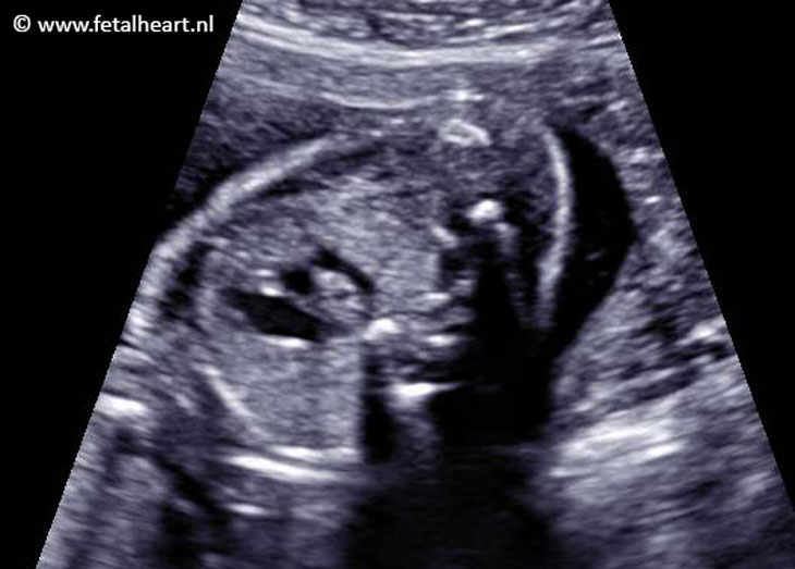
3VTV: ductal arch and aortic arch run both at the left side of the trachea.
At the roght side of the trachea a vessel is visible, this is the vena azygos entering the vena cava superior.
At the roght side of the trachea a vessel is visible, this is the vena azygos entering the vena cava superior.
Normal aortic arch with forward flow.
Final image showing a left pulmonary vein entering the left atrium.