You are here:
DORV-Fallot case 1
123456789
Transverse plane through the thorax at the level of the 4 chamber view.
However, only one ventricle is visible, containing the moderatorband.
At the left side a very small residual ventricle is visible.
The left atrium is smaller than the right atrium.
However, only one ventricle is visible, containing the moderatorband.
At the left side a very small residual ventricle is visible.
The left atrium is smaller than the right atrium.
Right ventricle with 1 unidentified outflow tract.
In this clip the foramen ovale and pulmonary veins entering the left atrium are clearly visible.
In this clip the foramen ovale and pulmonary veins entering the left atrium are clearly visible.
Clip with color Doppler.
In blue the inflow in the ventricle.
In red the aorta.
At the end of the clip is visible that this large vessel continues into the aortic arch (becoming blue because direction turns towards the spine).
The crossing pulmonary artery is visible in a blue-white signal (aliasing).
This is increased velocity across the pulmonary valve with in red the reversed flow in the ductus arteriosus.
Transverse plane in the upper part of the thorax at level of the 3VTV.
The duct is a bit tortuous and red (reversed filling).
The duct is a bit tortuous and red (reversed filling).
Aortic arch, arising too anteriorly.
The ascending aorta is a bit too large.
The ascending aorta is a bit too large.
Aortic arch with color Doppler.
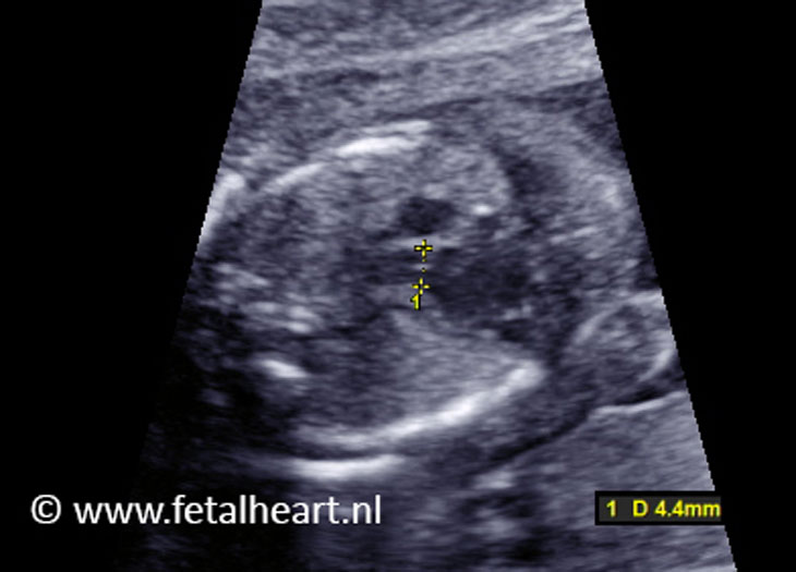
Aortic valve annulus.
4.4.mm is too wide for 20 weeks’gestational age.
4.4.mm is too wide for 20 weeks’gestational age.
Clip containing the ventricle and both outflow tracts.
The foramen ovale is clearly visible, as well as the pulmonary veins entering the left atrium.
Furthermore the pulmonary artery is clearly visible (although very small) from this insonation angle.
The foramen ovale is clearly visible, as well as the pulmonary veins entering the left atrium.
Furthermore the pulmonary artery is clearly visible (although very small) from this insonation angle.
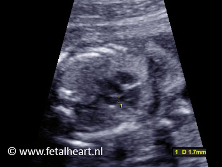
Measurement of the pulmonary valve.
1.7 mm is too small for 20 weeks’ gestational age.
1.7 mm is too small for 20 weeks’ gestational age.