You are here:
HLHS AoA case 1
12345
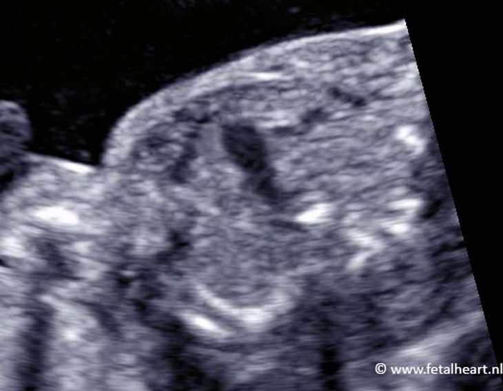
Plane of the 4 chamber view.
Only 1 ventricle is visible, slightly hypertrophic.
The left ventricle is hardly visible.
The thickened papillary muscle in the centre of the ventricle is mimicking the ventricle septum.
Note the atriumseptum, positioned too leftwards.
Only 1 ventricle is visible, slightly hypertrophic.
The left ventricle is hardly visible.
The thickened papillary muscle in the centre of the ventricle is mimicking the ventricle septum.
Note the atriumseptum, positioned too leftwards.
3 vessel view.
The ascending aorta is visible but too small.
The ascending aorta is visible but too small.
3VTV: the red flow is the small aortic arch with reversed filling.
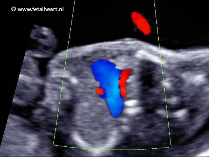
Ductal and aortic arch.
The antegrade flow in the duct is blue.
The aortic arch, cranial of the duct, is in red, indicating reversed filling.
The antegrade flow in the duct is blue.
The aortic arch, cranial of the duct, is in red, indicating reversed filling.
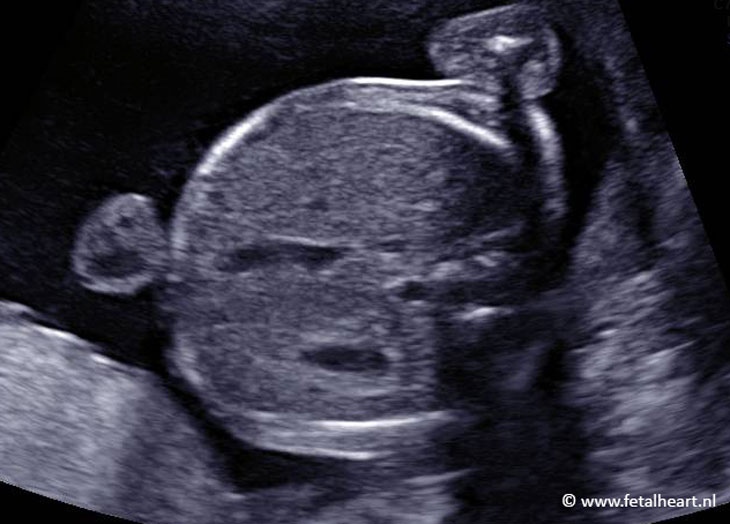
Normal adominal situs.