You are here:
HLHS AoS case 2
1234567
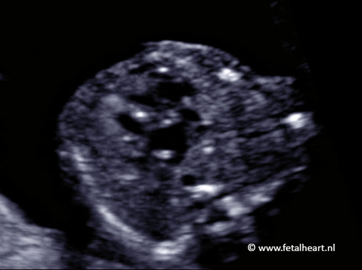
4 chamber view.
The left ventricle does not contract, has a global shape and shows myocardial hypertrophy (thickening).
Note the bright hyperechogenic dots at the ventricular septum (fiboelastosis).
The atrial septum is thickened and does not show the opening of the foramen ovale.
The left ventricle does not contract, has a global shape and shows myocardial hypertrophy (thickening).
Note the bright hyperechogenic dots at the ventricular septum (fiboelastosis).
The atrial septum is thickened and does not show the opening of the foramen ovale.
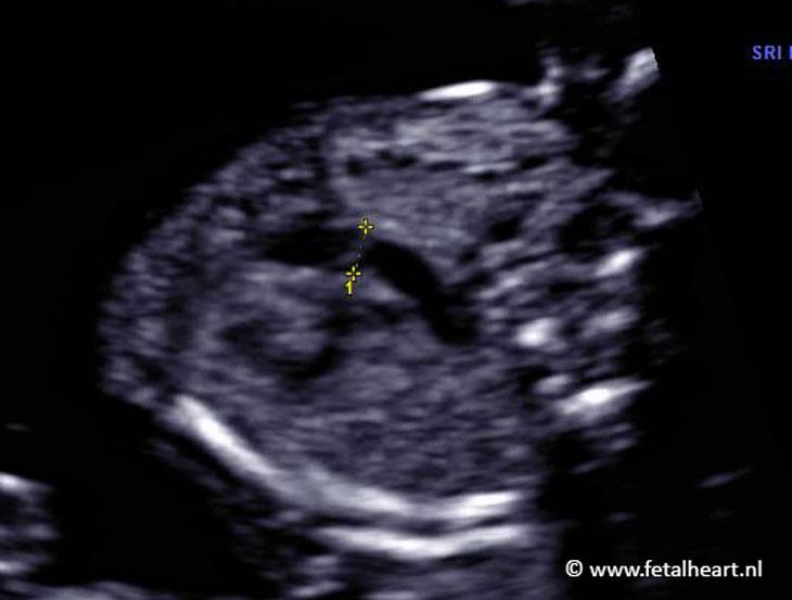
Still of the 4 chamber view.
Note the difference in size of both ventricles, although the left ventricle still shows a reasonable lumen.
The ventricle wall is thickened.
Note the difference in size of both ventricles, although the left ventricle still shows a reasonable lumen.
The ventricle wall is thickened.
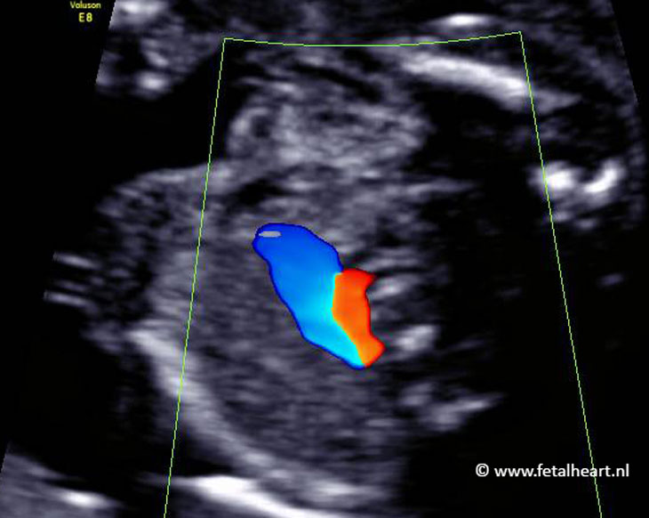
Color Doppler recordings shows that hardly any blood is entering the left ventricle.
Note the very clear signals of the pulmonary veins.
This is because the pressure in the pulmonary venous system is increased.
Note the very clear signals of the pulmonary veins.
This is because the pressure in the pulmonary venous system is increased.
Measurement of the pulmonary artery.
Size is a bit too large for 20 weeks gestational age.
Size is a bit too large for 20 weeks gestational age.
Still of the 3VTV: the red signal is the reversed flow in the aortic arch.
This clip shows flow back and forward in the pulmonary vein.
This is a sign of increased pulmonary venous pressure.
Flow across the foramen ovale is also absent.
This is a sign of increased pulmonary venous pressure.
Flow across the foramen ovale is also absent.
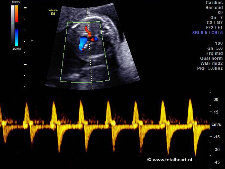
Pulsed wave Doppler signal of the pulmonary vein.
The peaks indicating reversed flow are abnormal and are associated with an adverse outcome.
The peaks indicating reversed flow are abnormal and are associated with an adverse outcome.