You are here:
PA-IVS case 1
1234567
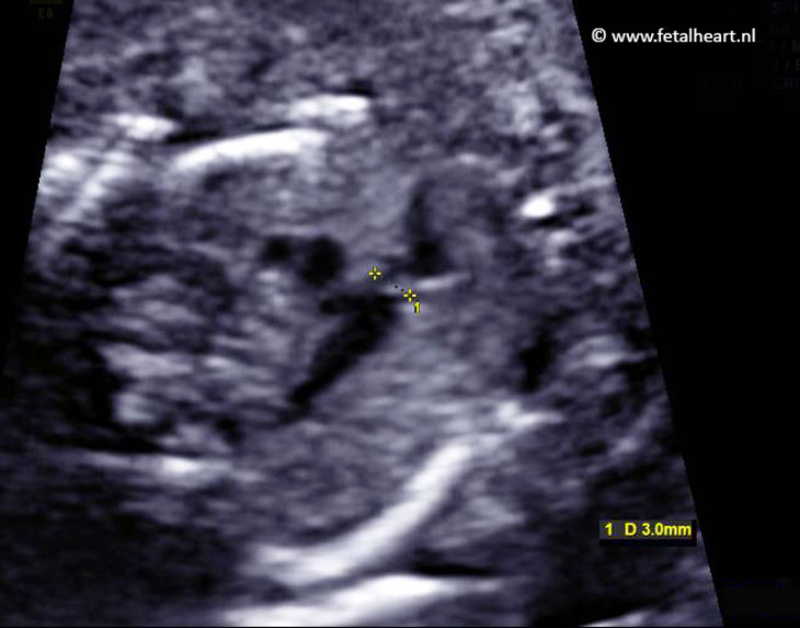
Asymmetrical 4 chamber view.
The right ventricle is very small and thickened.
The right ventricle is very small and thickened.
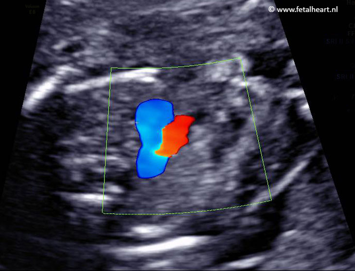
Color Doppler recording of the 4 chamber view.
Inflow in the right ventricle is minimal, with massive regurgitation over the tricuspid valve (red whirlpool).
Inflow in the right ventricle is minimal, with massive regurgitation over the tricuspid valve (red whirlpool).
Normal left ventricular outflow tract.
Note the non-contracting right ventricle.
Note the non-contracting right ventricle.
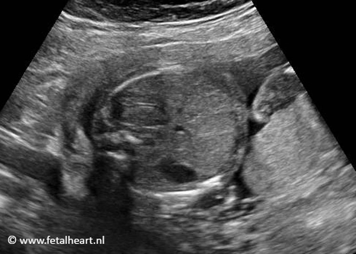
3 vessel view.
The pulmonary valve annulus is quite normal.
The fact that the valve is not opening is not visible in stills, you need real time imaging for that.
The pulmonary valve annulus is quite normal.
The fact that the valve is not opening is not visible in stills, you need real time imaging for that.
3VTV: the ductal arch is red, indicating reversed flow.
This is a sign of severe stenosis or atresia of the valve.
This is a sign of severe stenosis or atresia of the valve.
Normal abdominal situs.
Aortic arch and ductal arch in 1 clip.
Aortic arch is red (forward flow), ductal arch is blue (reversed flow).
Aortic arch is red (forward flow), ductal arch is blue (reversed flow).