You are here:
Unbalanced AVSD case 1
12345678
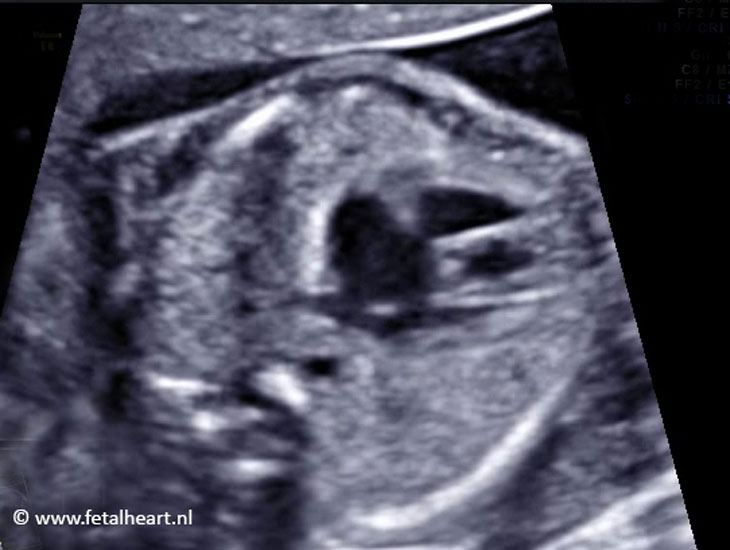
Asymmetrical 4 chamberview.
Left ventricle is smaller than the right ventricle.
Crux is absent.
The AV-valves are aligned and a bit bdomed.
Left ventricle is smaller than the right ventricle.
Crux is absent.
The AV-valves are aligned and a bit bdomed.
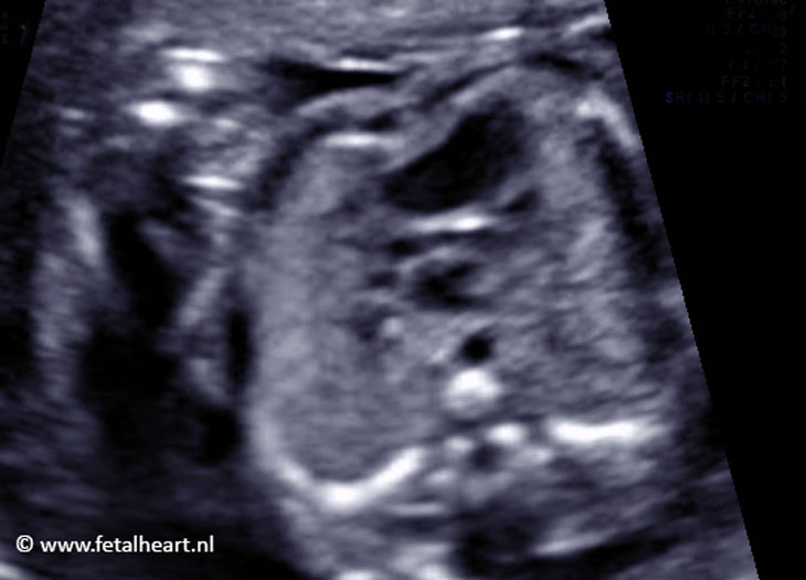
Asymmetrical 4 chamber view.
In the slow-motion part of the clip is the common AV-valve visible.
Crux is absent.
The pulmonary veins, entering the left atrium are clearly visible.
In the slow-motion part of the clip is the common AV-valve visible.
Crux is absent.
The pulmonary veins, entering the left atrium are clearly visible.
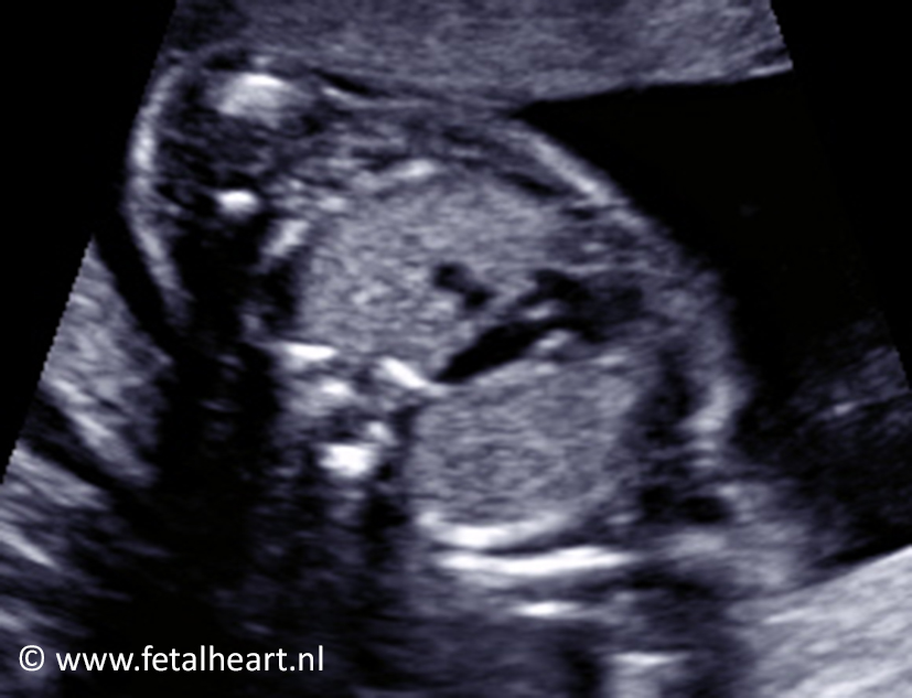
Color Doppler recording of the inflow of the ventricles.
The left ventricle receives less blood compared to the right ventricle.
Just beneath the common AV-valve is flow across the relatively small VSD visible.
The left ventricle receives less blood compared to the right ventricle.
Just beneath the common AV-valve is flow across the relatively small VSD visible.
Color Doppler recording of the flow across the atrial septum defect.
Still of the 4 chamber view.
The defect in the septum primum is clearly visible.
The defect in the septum primum is clearly visible.
Left ventricle outflow tract.
The aorta is small.
The aorta is small.
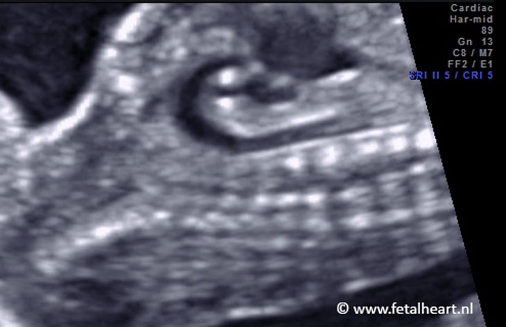
Three vessel view.
The ascending aorta is small compared to the pulmonary trunk.
The ascending aorta is small compared to the pulmonary trunk.
Aortic arch.
The ascending part is small, but the arch is well developed.
The ascending part is small, but the arch is well developed.