You are here:
VSD case 1
123456
Normal 4 chamber view at 19 weeks’ gestational age.
Echogenic focus in the left ventricle.
Echogenic focus in the left ventricle.
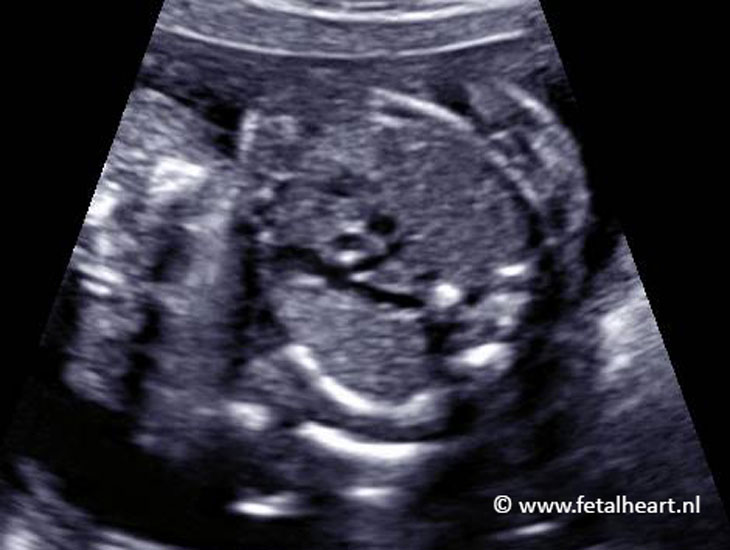
At 28 weeks’ gestational age. The VSD in the septum is clearly visible.
Left ventricle outflow tract.
VSD is just beneath the aorta.
VSD is just beneath the aorta.
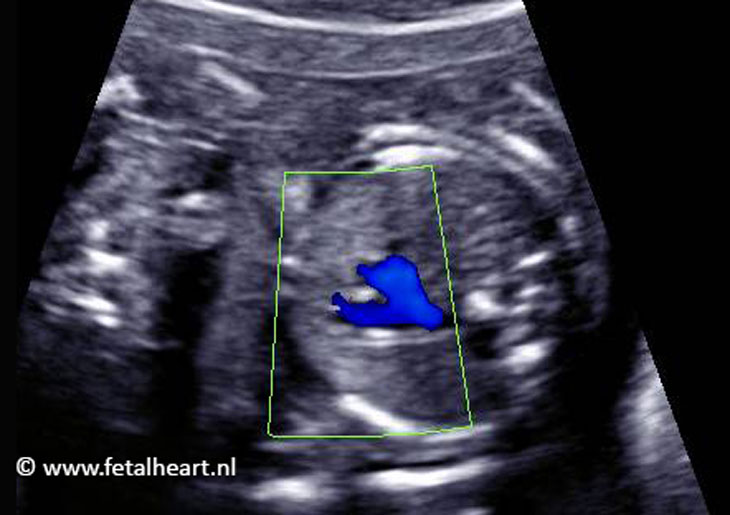
With color doppler a jet is visible beneath the aortic valve.
Blue and red flow is visible across the VSD, thus blood is traveling from the right to the left ventricle and vice versa.
This is a normal phenomenon in fetal VSDs.
Blue and red flow is visible across the VSD, thus blood is traveling from the right to the left ventricle and vice versa.
This is a normal phenomenon in fetal VSDs.

Normal 3 vessel view.

Normal 3VTV.
