You are here:
Ebstein case 1
12345678
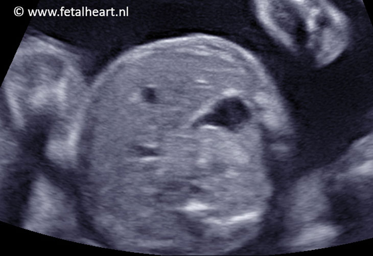
4 chamber view.
The ventricles are symmetrical.
Cardiac axis normal, crux is visible and appears normal.
The atria are not equally sized, the right atrium is bigger than the left one.
The attachment of the tricuspid valve to the ventricular septum is not strikingly abnormal from this insonation angle.
The ventricles are symmetrical.
Cardiac axis normal, crux is visible and appears normal.
The atria are not equally sized, the right atrium is bigger than the left one.
The attachment of the tricuspid valve to the ventricular septum is not strikingly abnormal from this insonation angle.
4 chamberview visualised from other insonation angle.
The enlarged right atrium is evident.
Note the mildly enlarged differential insertion.
The enlarged right atrium is evident.
Note the mildly enlarged differential insertion.
Apical 4 chamberview.
It looks like there are 2 AV-valves in the right ventricle: this is caused by the oblique positioned tricuspid valve in the ventricle.
The annulus is at normal level, the septal leaflet of the tricuspid valve is attached very apically, half-way down in the ventricle. This is very evident in the slow-motion part.
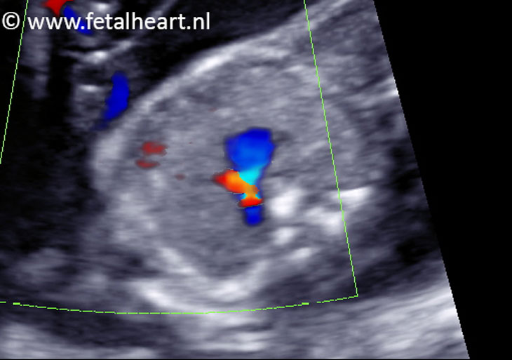
Color Doppler recording of the 4 chamber view.
An enormous jet across the AV-valve is visible (red whirlpool), indicating leakage.
An enormous jet across the AV-valve is visible (red whirlpool), indicating leakage.
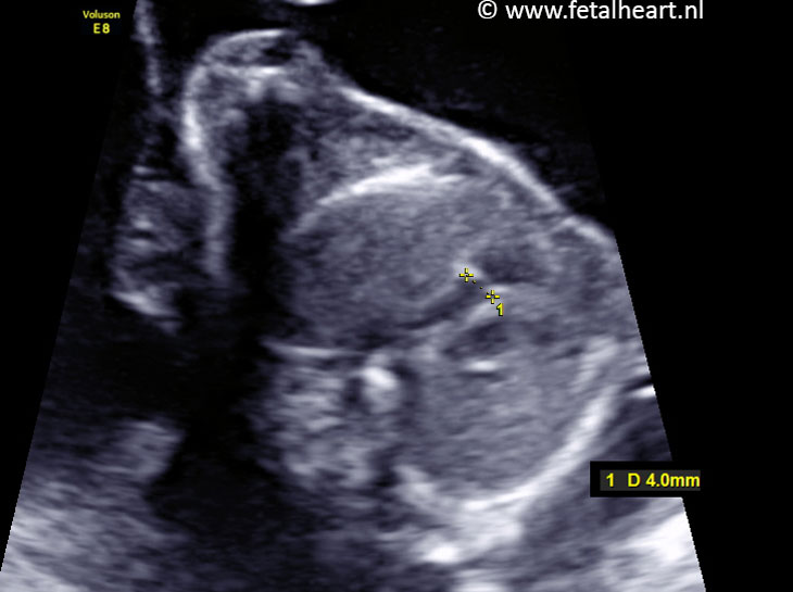
Measurement of the pulmonary trunk annulus.
Size within normal range.
Size within normal range.
3VTV in color Doppler.
Reversed flow in arterial duct, indicating functional or anatomic pulmonary atresia, which is a frequently encountered finding in Ebstein.
Reversed flow in arterial duct, indicating functional or anatomic pulmonary atresia, which is a frequently encountered finding in Ebstein.
Normal abdominal situs.
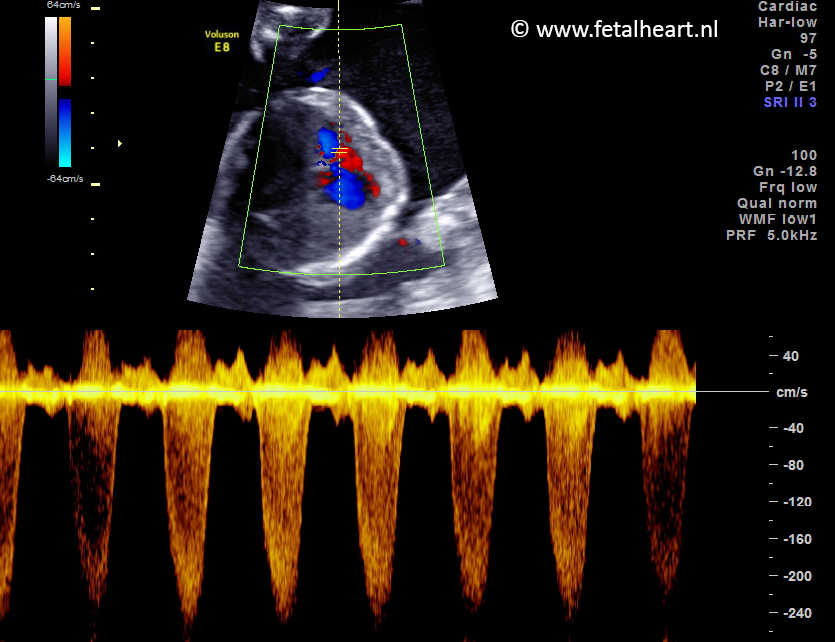
Pulsed wave recording of the massive leakage of the tricuspid valve.