You are here:
AoS case 2
123456789
Normal 4 chamberview.
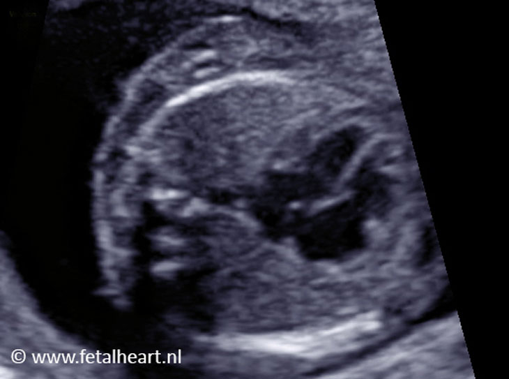
Still of the four chamber view.
No obvious anomalies in this image.
The foramen ovale valve is illustrative in this image.
No obvious anomalies in this image.
The foramen ovale valve is illustrative in this image.
Still of the 4 chamber view, tilting towards the left ventricle outflow tract.
Pulmonary veins entering the left atrium are clearly visible.
Pulmonary veins entering the left atrium are clearly visible.
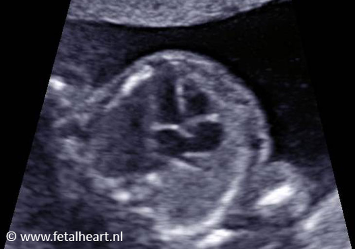
Left ventricular outflow tract.
Aortic valve is thickened.
The ascending aorta is widened after the valve (post-stenotic dilatation).
Aortic valve is thickened.
The ascending aorta is widened after the valve (post-stenotic dilatation).
Same as 4, bit from a different insonation angle.
Left ventricular outflow tract.
Aortic valve is thickened.
The ascending aorta is widened after the valve (post-stenotic dilatation).
Left ventricular outflow tract.
Aortic valve is thickened.
The ascending aorta is widened after the valve (post-stenotic dilatation).
3 vesselview.
Normal pulmonary trunk with lateral branching.
The ascending aorta is widened.
Normal pulmonary trunk with lateral branching.
The ascending aorta is widened.
3VTV: both arches show normal forward flow.
The color is different because of the position of the fetus.
The forward direction of the ductal arch is downwards, thus blue.
The forward direction in the aortic arch is upwards in the image, thus red.
The color is different because of the position of the fetus.
The forward direction of the ductal arch is downwards, thus blue.
The forward direction in the aortic arch is upwards in the image, thus red.
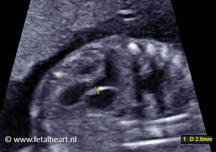
Measurement of the aortic annulus: normal size for 20 weeks’ gestational age.
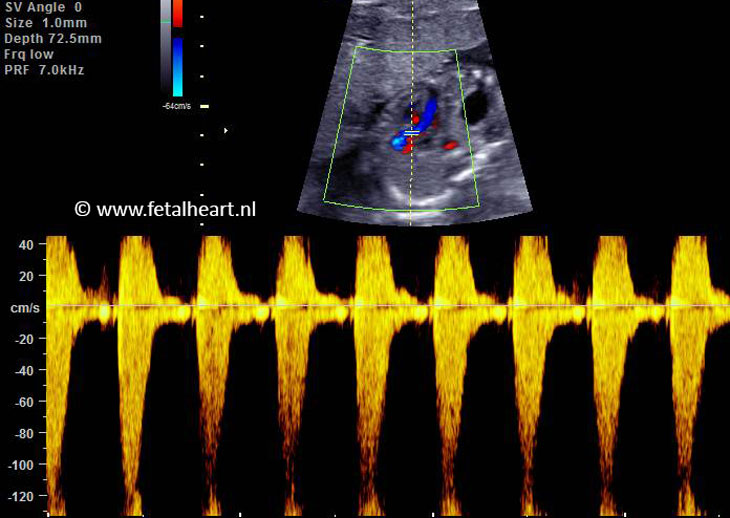
Increased velocities across the aortic valve, indicating aortic valve stenosis..