You are here:
Fallot case 1
12345678
More of less normal 4 chamber view.
The cardiac axis is abnormal however, (axis too large).
Differential insertion absent.
Note the difficulty that the examiner experiences in obtaining the 4 chamber view.
The cardiac axis is abnormal however, (axis too large).
Differential insertion absent.
Note the difficulty that the examiner experiences in obtaining the 4 chamber view.
Left ventricle outfow tract.
Overriding aorta is clearly visible.
Overriding aorta is clearly visible.
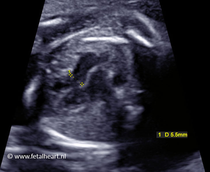
Image of measurement of the aortic annulus.
5.5 mm is too large for 20 weeks’ gestational age.
5.5 mm is too large for 20 weeks’ gestational age.
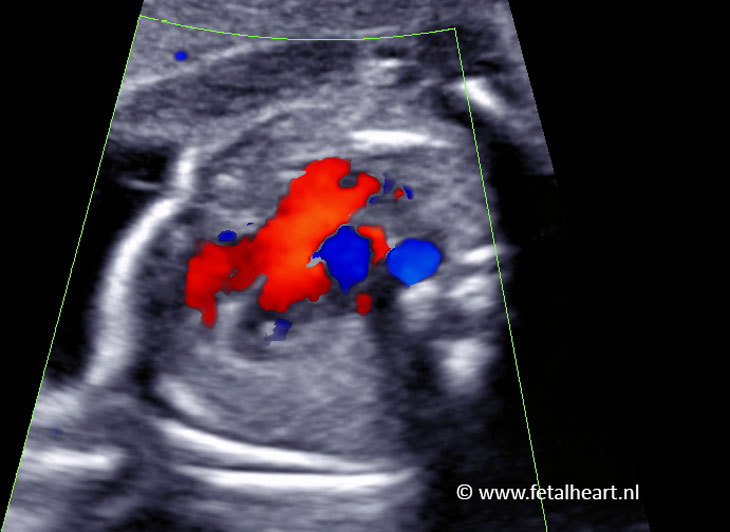
Still with color Doppler.
Aorta receives flow from the right and left ventricle.
Aorta receives flow from the right and left ventricle.
Sweep containing aorta and right ventricle outflow tract.
The pulmonary trunc is very small.
The pulmonary trunc is very small.
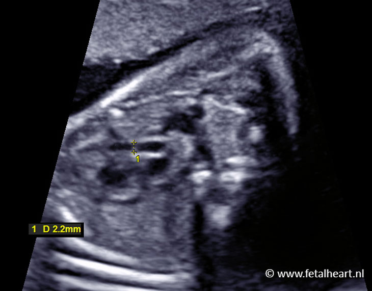
Measurement of the pulmonary trunc annulus.
2.2 mm is too small for 20 weeks’ gestational age.
2.2 mm is too small for 20 weeks’ gestational age.
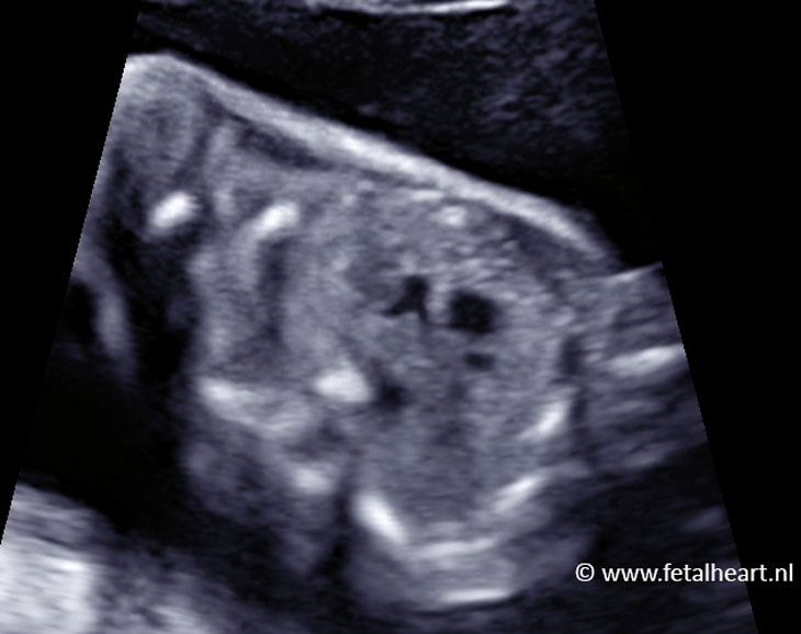
Three vessel view.
Aorta is large and positioned too anteriorly.
Pulmonary trunc is too small.
Aorta is large and positioned too anteriorly.
Pulmonary trunc is too small.
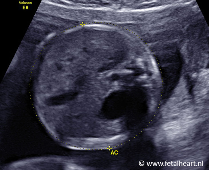
Normal transverse plane abdomen.