You are here:
Fallot case 2
12345678
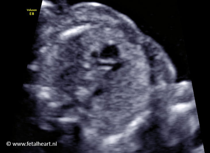
Normal 4 chamber view.
Differential insertion is absent (AV valve attach to the septum at the same level).
Differential insertion is absent (AV valve attach to the septum at the same level).
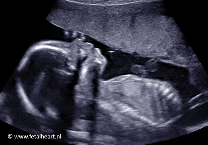
Clip with a sweep from 4 chamber view to aortic arch.
4 chamber view is normal (but with absent differential insertion), VSD is clearly visible (note the white spot on the top of the septum).
The aorta arises too much towards the right ventricle and overrides the VSD.
The pulmonary trunc is very tiny.
4 chamber view is normal (but with absent differential insertion), VSD is clearly visible (note the white spot on the top of the septum).
The aorta arises too much towards the right ventricle and overrides the VSD.
The pulmonary trunc is very tiny.
Still of the 3 vesselview.
Aorta is too large and is positioned too anteriorly.
Pulmonary trunc is very small, but the lateral branching is visible.
Aorta is too large and is positioned too anteriorly.
Pulmonary trunc is very small, but the lateral branching is visible.
Left ventricle outflow tract.
Aorta is not in alignment with the septum.
The walls of the aorta are not clearly visible in this projection.
Aorta is not in alignment with the septum.
The walls of the aorta are not clearly visible in this projection.
Aortic and ductal arch.
Aorta arises too anteriorly.
Underneath the aortic arch a red jet is visible: this is reversed flow in the ductal arch.
Aorta arises too anteriorly.
Underneath the aortic arch a red jet is visible: this is reversed flow in the ductal arch.
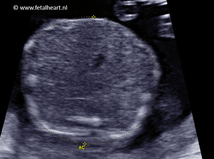
Transverse plane fetal abdomen: stomach not visible.
Face profile: absent nasal bone.
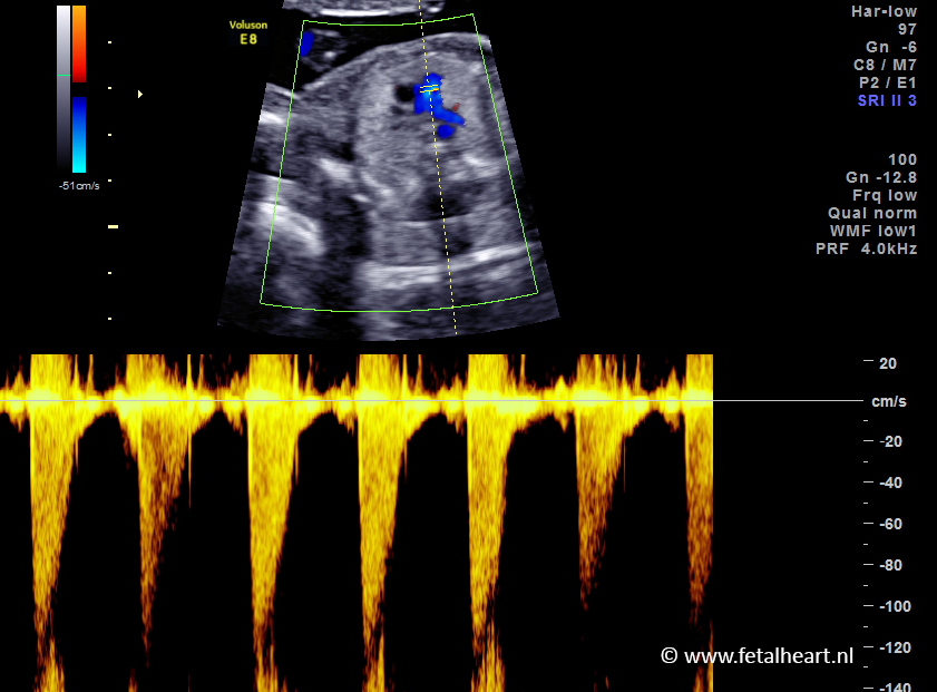
Pulsed wave Doppler across the pulmonary valve: increased velocities indicating severe stenosis.