You are here:
TGA case 2
123456789
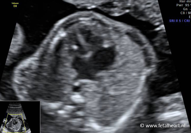
Normal 4 chamber view.
The atrial septum seems a bit thickened, but the upcoming images show no abnormalities in the trial septum.
The atrial septum seems a bit thickened, but the upcoming images show no abnormalities in the trial septum.
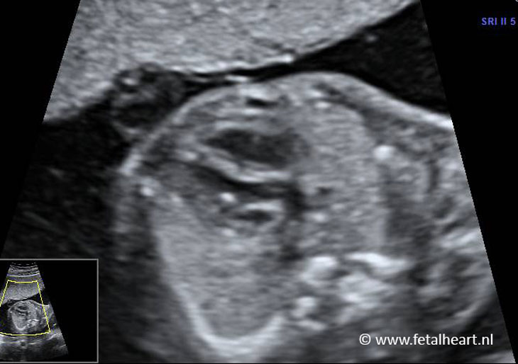
Apical insonation of the 4 chamber view.
Pulmonary veins entering the left atrium are clearly visible.
Left ventricle contains a golfball.
Pulmonary veins entering the left atrium are clearly visible.
Left ventricle contains a golfball.
Left ventricular outflow tract (LVOT): mimicking an aorta, but the course is abnormal.
The vessel bends leftwards immediately after the valve, instead of the normal course more rightwards.
The vessel bends leftwards immediately after the valve, instead of the normal course more rightwards.
Still of the LVOT similar as 3.
Abnormal course of the vessel bending leftwards.
Abnormal course of the vessel bending leftwards.
Clip illustrating the abnormal course of the LVOT, little bit more cranial is the outflow tract of the right ventricle visible.
RVOT had an abnormal course as well, too much rightwards.
RVOT had an abnormal course as well, too much rightwards.
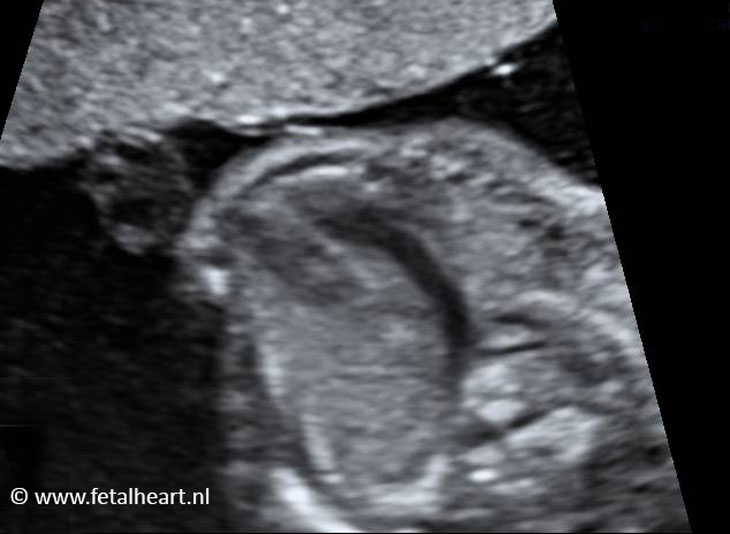
Image of the abnormal course of the vessel leaving the right ventricle.
Clip showing the parallel course of the great vessels.
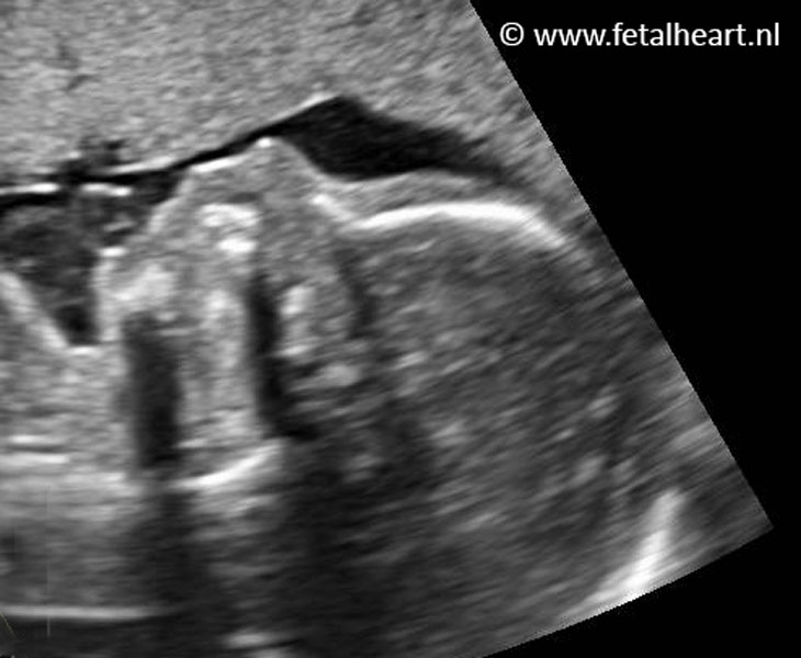
Aortic arch and ductal arch with color Doppler.
Most cranial arch is aortic arch, which starts directly behind the sternum, which is abnormal.
Most cranial arch is aortic arch, which starts directly behind the sternum, which is abnormal.
Normal profile of the face.