You are here:
TA case 2
123456
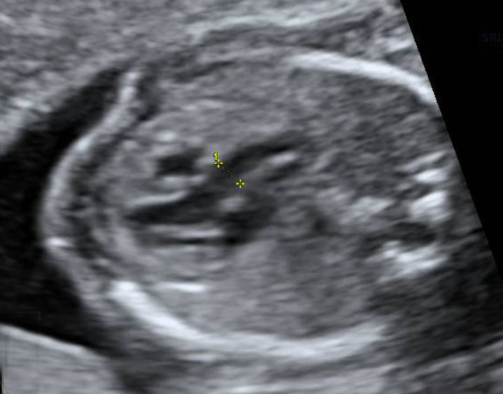
Asymmetrical 4 chamber view, with a very small right ventricle.
The left ventricle shows the typical morphological features of the left ventricle: papillary muscles attach to the free ventricle wall and continuity between mitral and aortic valve.
A large VSD is present.
The foramen ovale opens to the left atrium.
Just above atrial septum a white line is visible, this is the propbably a part of the ascending aorta.
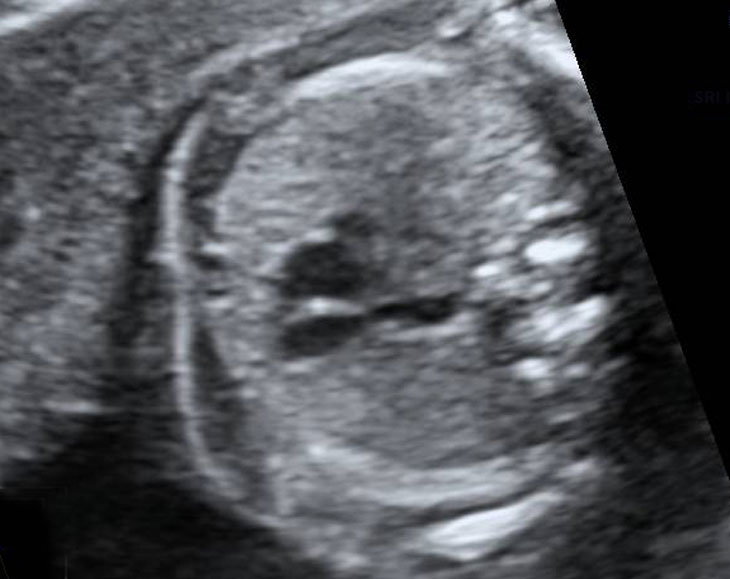
From this insonation angle is clearly visible that the AV-junction is closed.
Dense tissue is present at the site where the tricuspid valve should be opening.
The VSD is clearly visible as well.
Dense tissue is present at the site where the tricuspid valve should be opening.
The VSD is clearly visible as well.
Normal left ventricle outflow tract.
Aorta has a normal size and is in alignment with the ventricle septum.
Aorta has a normal size and is in alignment with the ventricle septum.
Clip with the outfllow tracts.
Aorta is normal, arising from the left ventricle.
The pulmonary trunk has an adequate size with a competent valve.
In tricuspid atresia pulmonary stenosis of atresia is not uncommon.
The size of the aorta is, however, slightly bigger that the pulmonary trunk.
Aorta is normal, arising from the left ventricle.
The pulmonary trunk has an adequate size with a competent valve.
In tricuspid atresia pulmonary stenosis of atresia is not uncommon.
The size of the aorta is, however, slightly bigger that the pulmonary trunk.
Three vessel view.
The subtle discrepancy in the outflow tracts is visible.
The subtle discrepancy in the outflow tracts is visible.
Still of the 3VV.
Aorta is bigger than the pulmonary trunk.
Aorta is bigger than the pulmonary trunk.