You are here:
AVSD case 2
1234567
4 chamber view.
Crux is absent.
Because of the small size of the VSD-component of this AVSD, the 4 chamber view looks relatively normal.
Crux is absent.
Because of the small size of the VSD-component of this AVSD, the 4 chamber view looks relatively normal.
Color Doppler shows the merged inflow centrally in the heart.
Duplex.
In the B image the common valve is visible with common inflow in the ventricles in red.
Crux is absent.
In the B image the common valve is visible with common inflow in the ventricles in red.
Crux is absent.
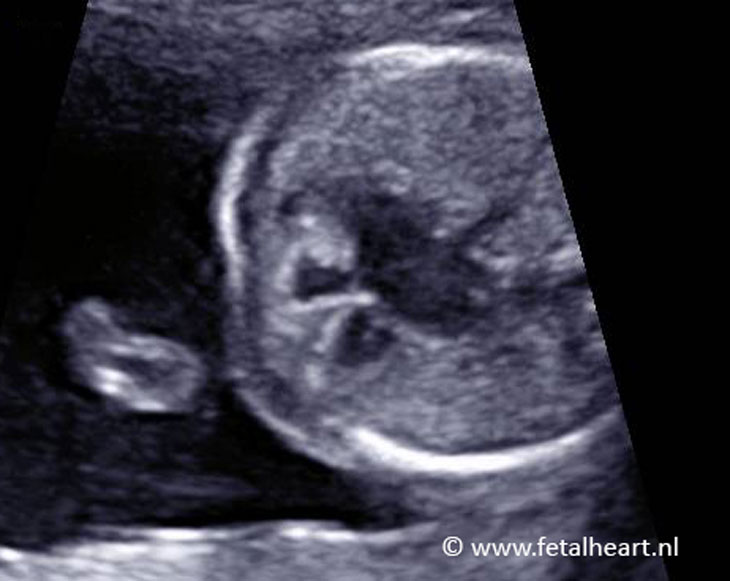
Still; absent differential insertion.
Absent crux.
Note the short length of the ventricle septum compared to the size of the atria.
Absent crux.
Note the short length of the ventricle septum compared to the size of the atria.
Normal spatial relaltionship of aortic arch and ductal arch (3VTV).
Normal abdominal situs.
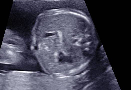
Ductal and aortic arch.
The aorta arises relatively anteriorly, this is caused by the AVSD.
The aorta arises relatively anteriorly, this is caused by the AVSD.