You are here:
AVSD case 3
1234567
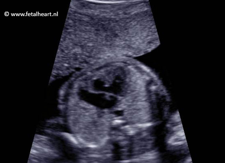
4 chamber view.
Ventricles are equally sized.
The crux is missing.
The common valve is difficult to see in this insonation angle.
Ventricles are equally sized.
The crux is missing.
The common valve is difficult to see in this insonation angle.
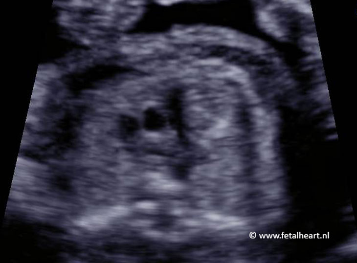
Apical insonation of the 4 chamber view.
AV valves form a straight line.
VSD is visible, recognizable by the white dot on the top of the septum.
AV valves form a straight line.
VSD is visible, recognizable by the white dot on the top of the septum.
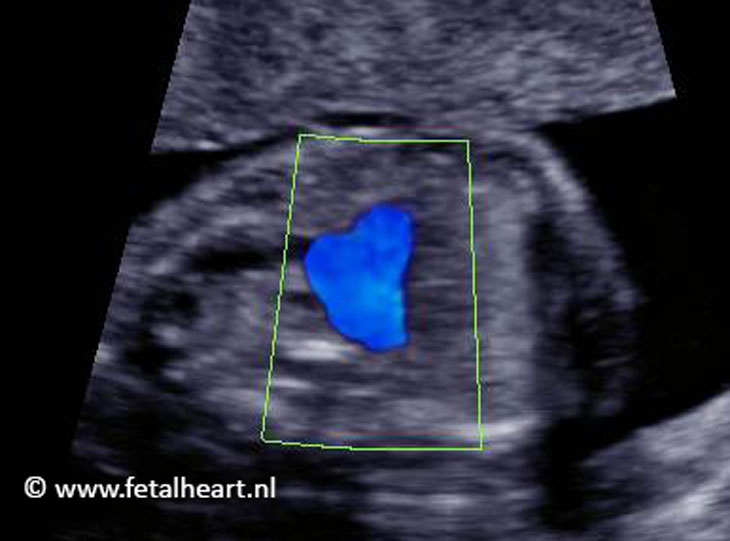
Clip of the apical 4-chamber view.
In slow motion the common AV-valve is clearly visible.
When scanning, you need the cine-loop function for visualise the common valve like this.
In slow motion the common AV-valve is clearly visible.
When scanning, you need the cine-loop function for visualise the common valve like this.
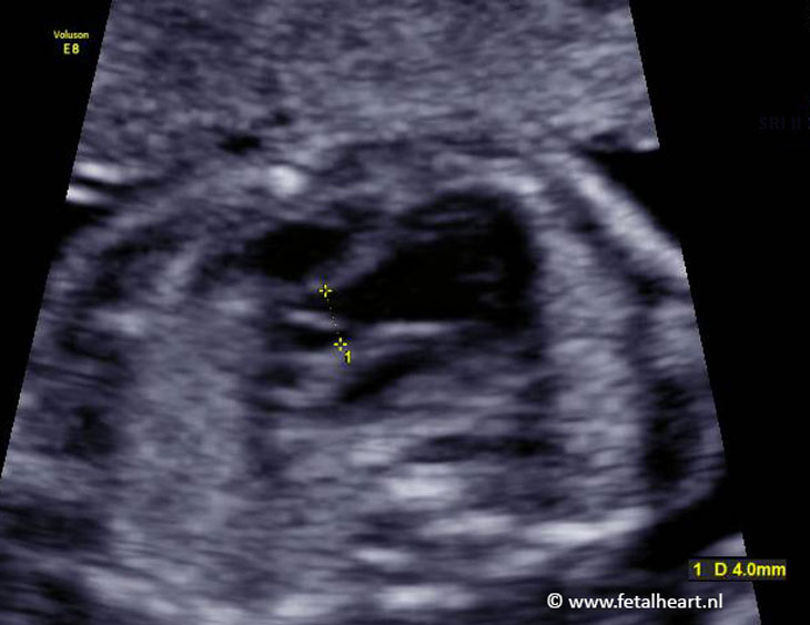
Image of measurement of aortic valve diameter.
4.0 mm is normal size for 22 weeks’ gestational age.
4.0 mm is normal size for 22 weeks’ gestational age.
Normal three vessel view.
Normal 3VTV.
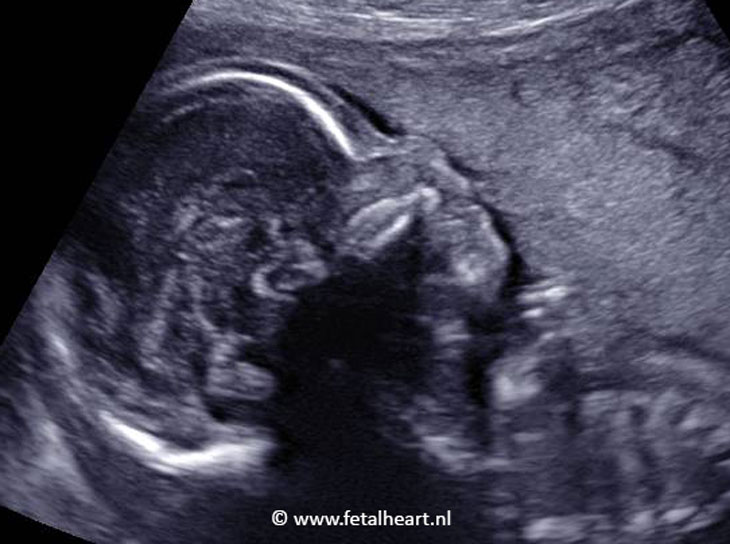
Profile of the face.
Absent nasal bone.
Combination of AVSD and absent nasal bone makes trisomy 21 very likely.
Absent nasal bone.
Combination of AVSD and absent nasal bone makes trisomy 21 very likely.